Association with Internal Diseases (For Professionals)
Association with Internal Diseases (For Professionals)
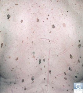
The rapid apparition of SK mostly when it is associated with acanthosis nigricans (35% of patients) and especially pruritus{Holdiness, 1988} should raise the suspicion about an internal malignancy. There is a big polemic about the existence of this rare sign called Leser-Trélat. The information concerning this sign is shown below.
The coexistence of internal cancer to the eruption of multiple SK in is called the sign of Leser-Trélat which has been described independantly by two European surgeons, Edmund Leser (1853-1916) from Germany {Leser, 1901 #222}, and Ulysse Trélat (1828-1890) separately at the beginning of the 20th century. Nevertheless, the sign of Leser-Trélat is not deserved by its authors, since the the description related the appearance of internal neoplasms with cherry angiomas (Campbell de Morgan spots).
In this sign, lesions crop on the whole body with a predilection for the back, arms and legs, face, abdomen, neck, axillary and inguinal folds {Yeh, 2000; Schwartz, 1996}. The cutaneous findings coincide with the progression of the internal neoplasm and may disappear after surgical or non surgical treatment and reappear with recurrence and metastasis {Pierson, 2003}.
Associated signs are sometimes found and several signs of malignancy have been reported associated such as Cowden’s disease {Aylesworth, 1982}, acquired ichtyosis {Hage, 1977}, acrokeratosis of Bazex {Rubisz-Brzezinska, 1983}, acquired hypertrichosis lanuginosa {Dingley, 1957; Jemec, 1986} tylosis {Millard, 1976}.
Of the more than 80 cases reported {Schwartz, 1996}, associated malignancies described include adenocarcinomas of the lung {Heaphy, 2000} renal cell carcinoma {Vielhauer, 2000}, colorectal cancer {Ginarte, 2001}, and most frequently reported adenocarcinoma of the stomach {Andreev, 1981; Stieler, 1987; Kameya, 1988; Sperry, 1980} {Tutakne, 1983; Hoffmann, 1989; Schwartz, 1978; Lindelof, 1992; Millard, 1976}. Other malignant conditions such as Sezary syndrome {Horiuchi, 1985} osteogenic sarcoma {Barron, 1992} and transitional cell carcinoma of the urinary bladder {Yaniv, 1994} have been reported. The prognosis for patients with the Leser-Trélat sign doesn’t exceed eleven months {Holdiness, 1986}.
The pathogenesis was first suspected in the first of two patients studied by Millar with tylosis who presented with the sign of Leser-Trélat {Millard, 1976}. This patient had high levels of immunoreactive growth hormone. Out of the second patient Ellis and Yates {Ellis, 1993} postulated that the sign of Leser-Trélat is the result of the action of a growth factor such as TGF alpha causing an eruption in a sensitized individual. TGF alpha exerts an action on EGF receptor which is present in vitro in peripheral lymphocytes in patients with SKs associated with lymphoma {Horiuchi, 1985} and in specimens of SKs in the second patient of Millar. Levels of EGF conversely are not increased. {Curry, 1980}.
Because of its rarity the existence of this sign has been questioned. Not only is the eruptive and acute apparition of SK found in conditions such as pityriasis rubra pilaris induced erythroderma {Schwengle, 1988}, pregnancy {Garcia, 1977} and HIV {Inamadar, 2003}, but it can also be found in the normal population {Lindelof, 1992}. We reported the case of an acute eruption of widespread SK following a heart transplant an immunosuppressive treatment with cyclosporine and prednisone {Hsu, 2005}.
Lindelof and his colleagues reviewed retrospectively 1752 patients wih a diagnostis of SK, then they figured out the yearly incidence of internal neoplasms which numbered 62 cases. 6 of these cases presented with eruptive SKs. A control group with the same characteristics of age and sex also had an identical incidence of eruptive SKs. This study thus rejected the validity of the Leser-Trélat sign assuming that both cancer and SK coexist because of their high prevalence. Nevertheless, the association of internal cancer and the acute cropping of SK has also been described in young patients in their twenties {Ellis, 1993; Barron, 1992}, thus opening the debate again.
Finally, we must point out that the diagnosis of the Leser-Trélat sign being based upon the abrupt onset of SKs associated with an internal neoplasia, précise quantification as to the length of the eruptive period and the number of SKs required for the diagnosis is lacking {Schwartz, 1996}.
This advice is for informational purposes only and does not replace therapeutic judgement done by a skin doctor.
Contributors:
Dr Christophe HSU – dermatologist. Geneva, Switzerland
Category : association avec maladies internes - Modifie le 10.7.2012Category : association with internal disease - Modifie le 10.7.2012Category : Kératose séborrhéique - Modifie le 10.7.2012Category : Seborrheic keratosis - Modifie le 10.7.2012Category : Seborrhoeic keratosis - Modifie le 10.7.2012Category : verrue séborrhéique - Modifie le 10.7.2012


