Dermoscopical Features (dermatoscopy) (For Professionals)
Seborrheic Keratosis = SK
Dermoscopical Features (dermatoscopy) (For Professionals)
– Hairpin vessels are long capillary rings which develop at the periphery of keratinising tumours {Kreusch, 1996}. Typically, many vessels bunch together and are surrounded with a white halo, thus giving a grape-like appearance {Braun, 2002}. This characteristic is more frequent in irritated SKs {Braun, 2002; Braun, 2002} {Stolz, 2002}. This sign is not specific, as melanomas can also display hairpin vessels.
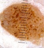
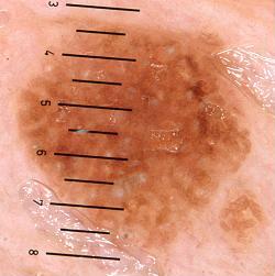
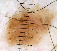
– Network-like pattern and fingerprinting : it doesn’t always resemble the pigmented network of benign melancytic lesions, which consist of grid-like {Menzies, 1996; Argenziano, 1998; Argenziano, 2000 ; Soyer, 2000; Kenet, 1993; Kreusch, 1991} {Stolz, 2002; Yadav, 1993} hyperpigmented lines surrounding hypopigmented zones. The lines of the network-like structures are much larger than those in the typical pigment network and may end abruptly at the periphery {Braun, 2002}. {Braun, 2002}The lines correspond to mild epidermal hyperplasia, hyperkeratosis and an increase of melanin in the keratinocytes {Schiffner, 2000}. The holes, which are much larger than in the melanocytic grid-like network, often correspond, as with fissures and comedo-like openings, to keratin-filled structures, not always to the tips of dermal papillae {Schiffner, 2000}.
– Moth-eaten borders {Schiffner, 2000} are truncated irregular borders seen in early SKs and solar lentigos.
– Wobble pattern {Braun, 2002}. On examining a skin lesion, it has been noticed that SK {Braun, 2000} adheres to the dermatoscope when the device is slightly moved horizontally and parallel to the skin surface. Moreover SKs have such a solid consistency that its image on the dermatoscope doesn’t change when the dermatoscope is moved.
– Sharp dermarcation {Braun, 2002} is present in nearly 90% of lesions in a study of 204 pigmented SKs .
– Some characteristics are more frequent than others. According to one study {Braun, 2002} of 203 pigmented SKs, the most frequently found lesions were milia-like cysts (135) and comedo-like openings (144 lesions), followed by hairpin vessels and fissures. 183 lesions were sharply demarcated. Nevertheless, the weakness of this study is that it aims pigmented SKs, not seborrheic keratosis as a whole. That means that the 94 network-like structures found are an overestimated figure.
According to our experience we evaluated the frequency of characteristics. The lesions were shown to us after being clinically diagnosed by clinicians as SK. The three most specific criteria for diagnosing SKs were sharp delineation, milia-like cysts and comedo-like openings. The presence of these three characteristics together is very specific. It diminishes in diagnostic power when one of these three features is absent.
Differential Diagnosis on dermoscopy
Differentiation between SK and melanoma is difficult in some cases when examined with a dermatoscope.
A retrospective study shows that out of 9204 cases clinically suggestive of SK, 61 (0,66%) turned out to be melanomas {Izikson, 2002}even though the differential diagnosis of melanoma had been evocated. More intriguing was the fact than in three of these 61 cases, seborrheic keratosis was the only suspected clinical diagnosis.
Argenziano et al (2003) {Argenziano, 2003} reported one case of a patient with a unique clinical lesion, suggestive of a SK; it was sharply demarcated but the patient had noticed in it a recent change in colour. On dermoscopical examination comedo-like openings and a homogenous network were present. There was a faint pigment network at the periphery and discrete pseudoreticular blue-gray structures around the comedo-like openings were found. Histological examination revealed a psoriasiform epidermal hyperplasia with keratin plugs but containing atypical melanocytes. Nests of melanocytes were found and melanophages were in the papillary dermis, the equivalent of blue-gray structure seen on dermoscopy. The diagnosis was melanoma in situ.
The conclusion of Argenziano (2003) {Argenziano, 2003} is that melanomas can occasionally present with certain features of SK (milia-like cysts, comedo-like openings) on dermoscopy even though he only described an isolated case.
Conversely SK can be diagnosed histologically after being dermoscopically suggested as being a melanocytic lesion. 10% of 402 lesions histologically classified as SK revealed a network pattern suggestive of a melanocytic lesion{De Giorgi, 2002}. Moreover, the most frequent found in seborrheic keratosis are sometimes not found; Hirata and al (2004) {Grichnik, 2004} report two cases of clinically pigmented lesions. No dermoscopical criteria for seborrheic keratosis was present except for globule-like structures, therefore a melanocytic lesion was suspected. Histological diagnosis revealed clonal type of SK.
This advice is for informational purposes only and does not replace therapeutic judgement done by a skin doctor.
Contributors:
Dr Christophe HSU – dermatologist. Geneva, Switzerland
Category : dermatoscopie - Modifie le 10.9.2012Category : dermatoscopy - Modifie le 10.9.2012Category : dermoscopical features - Modifie le 10.9.2012Category : dermoscopie - Modifie le 10.9.2012Category : dermoscopy - Modifie le 10.9.2012Category : Kératose séborrhéique - Modifie le 10.9.2012Category : Seborrheic keratosis - Modifie le 10.9.2012Category : Seborrhoeic keratosis - Modifie le 10.9.2012Category : verrue séborrhéique - Modifie le 10.9.2012


