Skin lesions in Crohn’s disease (for professionals)
Dr Christophe HSU – dermatologist. Geneva, Switzerland
- Crohn’s disease is an inflammatory disease of the digestive tract. It is clinically characterized by abdominal pain, bloody stools…Histologically, structures called granulomas are found in the inflammatory affected areas. We are interested in the skin problems which can be associated with this condition because we have observed therapeutical similatrities to treat this condition as well as psoriasis and hidradenitis suppurativa. This being the biologics.
The direct manifestations of Crohn’s disease are:
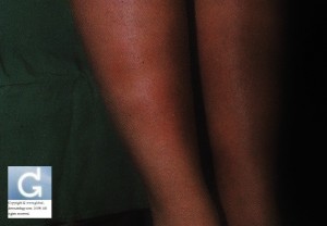
- Erythema Nodosum
- prodromal phase: lasts 3 to 6 days and is accompanied by pain and fever.
- induction phase: appearance of painful deep nodules over 1 to 2 days. Pain is alleviated by lying down.
- regressive phase: disappears in 10 days without leaving scars.
- Pyoderma Gangrenosum or more generally other neutrophilic dermatoses (defined by neutrophilic infiltrate with no infection)
- Sweet’s syndrome (Sweet’s syndrome in a patient with Crohn’s disease: a case report. Mustafa NM, Lavizzo M. J Med Case Reports. 2008 Jun 28;2:221.)
- Disseminated pustular eruption
- psoriasis (we have put it here because psoriasis in its pustular form is considered by some as a neutrophilic dermatosis)
- aseptic abscess ([Cutaneous aseptic abscesses, manifestations of neutrophilic diseases]Carvalho P, Cordel N, Courville P, Leloet X, Heron F, Lauret P, Joly P. Ann Dermatol Venereol. 2001 May;128(5):641-3. French.).
- Sneddon Wilkinson syndrome (Subcorneal pustulosis)(Subcorneal pustular dermatosis in a patient with Crohn’s disease. Delaporte E, Colombel JF, Nguyen-Mailfer C, Piette F, Cortot A, Bergoend H. Acta Derm Venereol. 1992 Aug;72(4):301-2.)
- Skin lesions located at or close to the abdomen, perineum or at a colostomy site.
- “stab like” linear fissures
- peripheral chronic perianal fissures
- papular and granulkar erosive lesions in the folds (some lesions at the inguinal-scrotal area can clinically mimic extramammary Paget’s disease: however biopsy shows granulomas).
- sometimes pseudocondylomatous lesions
- lesions are preferrentially located in areas of trauma (pathergy)
- Metastatic Crohn’s disease
- fissuring nodules or granulomes in the folds
- most often, these lesions are located on the trunk and genital area, but lesions have been described on the face and on the ears.
- More rarely:
- Oral lesions: pseudo-aphtous ulcers
- lesions on the lips looking like macrocheilitis of Melkersson-Rosenthal
- SAPHO syndrome: 6 patients (5%) out of 120 in this retrospective study had Crohn’s disease (SAPHO syndrome: a long-term follow-up study of 120 cases. Hayem G, Bouchaud-Chabot A, Benali K, Roux S, Palazzo E, Silbermann-Hoffman O, Kahn MF, Meyer O. Semin Arthritis Rheum. 1999 Dec;29(3):159-71.)
- Porokeratosis (Porokeratosis and Crohn’s disease. Morton CA, Shuttleworth D, Douglas WS. J Am Acad Dermatol. 1995 May;32(5 Pt 2):894-7.)
- Palmar erythema
- Finger clubbing
- Acquired epidermolysis bullosa
- Linear IgA dermatosis
- Because of Crohn’s disease, there is a subsequent increased risk of cancers (prolonged inflammation) and nutrition-deficency linked skin eruptions (due to malabsorption).
- Increased risk of cancer. Skin cancers like squamous cell carcinoma (Increased risk for non-melanoma skin cancer in patients with inflammatory bowel disease. Long MD, Herfarth HH, Pipkin CA, Porter CQ, Sandler RS, Kappelman MD. Clin Gastroenterol Hepatol. 2010 Mar;8(3):268-74. Epub 2010 Jan 16.) are more frequent and skin metastases of internal cancers can appear on the skin (pancreas, liver, lung, prostate, testicles, kidney, endocrinial tumours, lymphoma, leukemia)(Cancer risks in Crohn disease patients. Hemminki K, Li X, Sundquist J, Sundquist K. Ann Oncol. 2009 Mar;20(3):574-80. Epub 2008 Sep 2.)
- Nutrition-deficiency linked skin eruptions. Theoretically, malaborption can lead to numerous skin eruptions but in practice acrodermatitis-periorificial dermatitis is the most often encountered. It is due to a deficiency of zinc and is more frequen in infanthood when deficiency in zinc, protein, biotine and essential fatty acids give rise to acrodermatitis-periorificial dermatitis accompanied with a failure to thrive (Acrodermatitis due to nutritional deficiency. Gehrig KA, Dinulos JG. Curr Opin Pediatr. 2010 Feb;22(1):107-12. Review.)
Bibliography from non-cited references: Dermatologie et infections sexuellement transmissible, 4e édition
This advice is for informational purposes only and does not replace therapeutic judgement done by a skin doctor.
Contributors:
Dr Christophe HSU – dermatologist. Geneva, Switzerland
Category : acrodermatite - Modifie le 06.1.2010Category : acrodermatitis - Modifie le 06.1.2010Category : carcinome spinocellulaire - Modifie le 06.1.2010Category : crohn métastatique - Modifie le 06.1.2010Category : Crohn's disease - Modifie le 06.1.2010Category : dermatite périorificielle - Modifie le 06.1.2010Category : erythema nodosum - Modifie le 06.1.2010Category : érythème noueux - Modifie le 06.1.2010Category : maladie de Crohn - Modifie le 06.1.2010Category : metastatic Crohn's - Modifie le 06.1.2010Category : periorificial dermatitis - Modifie le 06.1.2010Category : porokératose - Modifie le 06.1.2010Category : porokeratosis - Modifie le 06.1.2010Category : pyoderma gangrenosum - Modifie le 06.1.2010Category : sapho syndrome - Modifie le 06.1.2010Category : squamous cell carcinoma - Modifie le 06.1.2010Category : syndrome de SAPHO - Modifie le 06.1.2010


