Morphea (Localized Scleroderma) (For Professionals)
Dr Christophe HSU – dermatologist. Geneva, Switzerland
Morphea is a disorder which is characterized by thickened skin lesions. This disorder results when there is excessive collagen deposition in the deeper part of the skin called the dermis and the fatty subcutaneous tissues of the skin. This skin disorder if often classified as generalized, plaque, or deep, depending on its presenting features and depth of tissue invasion.
Morphea is often compared with another similar disorder, systemic sclerosis. Systemic sclerosis also presents as skin thickening however there are additional features such as:
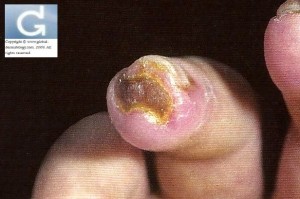
-thickening of the skin in the fingers and toes (sclerodactyly)(acrosclerosis) often resulting in ulcers
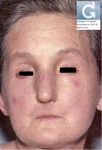
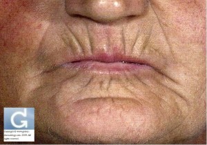
-mask-like facial features with fibrosis of the lips (thinning, pursed lips) and the perioral region resulting in a small mouth.
-discoloration of the fingers, toes and other areas (Raynaud phenomenon)
-changes in the nail’s appearance, visible dilatation of the blood vessels on the skin and/or the mucosal surface. (Dilatation (dilation) of blood vessels under the nail visible with the dermatoscope)
-involvement of other areas of the body (joints, bones…)
The severity of systemic sclerosis is determined according to the classification of Barnett with lesions being more and more proximal as the stage (I to III) increases and that the prognosis worsens (internal involvment).
There is also a more benign form of systemic sclerosis called CREST. This form is characterized by skin Calcinosis, Raynaud’s phenomenon, Esophageal Dysmotiliy, Sclerodactyly and cutaneous Telangiectasia.
Systemic Sclerosis can lead to complications related to fibrosing of the internal organs such as the heart (arrtythmias), the esophagous (dysmotility), pulomnary fibrosis (dyspnoea), kidney fibrosis (renal failure)…
Why does Morphea occur?
The main reason why this skin disorder occurs is overproduction is collagen by cells in the fibrous tissue called fibroblasts. This results after there is injury to the lining of the blood vessels, a lesion happening in immune disorders which is brought about by a reaction from foreign bodies or the body’s own cell, inflammation and an unregulated production of collagen.
Diagnosis of morphea is made on the clinical and histopathological (dermatopathology, skin microscopy) features with sometimes accompanying blood test anomalies with presence of autoantibodies such as ANA (Antinuclear antibodies). Systemic sclerosis on the other hand is also characterized by the presence of other autoantibodies such as anticentromere autoantibodies and scl-70 (DNA topoisomerase 1).
Clinical presentation
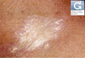
Clinically, skin lesions appear as whitish or yellowish dry skin. There are plaques which may appear slightly depressed, hard and oval in shapes that have whitish centers surrounded by purple or pinkish halos. You will often see these skin lesions on the trunk, however they can appear anywhere in the body (limbs…).
Morphea can be classified according to depth of tissue involvement. The basic classification is the following: (a) plaque type, (b) generalized type, (c) linear type, and (d) deep type:
a) The plaque type is the most common type of Morphea which affects the skin and spares the underlying tissues. These skin lesions often fade even without treatment within 3 to 5 years after onset.
b) The second type is the generalized type wherein skin lesions are very widespread. In this type, the skin lesions are hard and dark and spread to other large parts of the body. The lesions may also spread to the underlying muscles, causing it to become very hard, tight and atrophied.
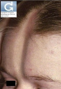
c) The third type is linear Morphea. This appears as a line of thickened skin which could affect bones and muscles underneath, to limit the movement of the affected muscles and joints. Linear Morphea occurs frequently in young children and manifests itself (when affected) as failure to grow one arm rapidly as expected. This type is often found in the arms, legs, and forehead (frontoparietal: “coup de sabre”) or in many other areas. It may occur on one side of the body only. It can be associated with hemiatrophy of the tongue.
d) The last type of Morphea is deep Morphea (atrophoderma of Pierini and Pasini). This lesion appears as ill-defines, hard plaques with a cobblestone appearance. It is best suspected when the groove sign is present: it appears as a depression along the course of a vein between two muscles or encompassing both muscles and veins. They are highly pigmented and are symmetric i.e. appears in both sides of the body.
In the hair and nails, involvement of any type can create permanent scarring alopecia.
In the pansclerotic type, the trunk and extermities (except fingertips and toes) are involved with the dermis, fat, fascia and bone being involved.]
The course of the disease is slowly progressive, but spontaneous remissions can occur.
Contributors:
Dr Christophe HSU – dermatologist. Geneva, Switzerland
Category : Localized Scleroderma - Modifie le 03.28.2012Category : Morphea - Modifie le 03.28.2012Category : morphée - Modifie le 03.28.2012Category : Sclérodermie - Modifie le 03.28.2012Category : Sclérodermie Localisée - Modifie le 03.28.2012Category : Systemic Sclerosis - Modifie le 03.28.2012
