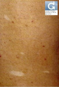
Differential diagnosis of hypomelanoses in children
Differential diagnosis of hypomelanoses in children (N. Van Geel (Ghent, Belgium))
Use a stepwise approach
step 1 history
- congenital or acquired
- stable or progressive
- other medical problems
- family history
step 2 clinical examination
- localisation (Woods light important especially in the localized form)
- number of lesions
- pattern (linear blaschkoid, segmental)
- hypomelanosis or amelanosis
- other problems (neurological, eyes)
step 3: Apply information of flowchart (differential diagnosis)
- if diffuse: presence or absence of ocular involvmenet
- ocular involvement: albinos group, also Griscelli syndrome, Chediak-Higashi syndrome
- no ocular involvement: think of Metabolic causes: Menkies syndrome, phenylcetonuria
- if localized
- depigmentation: waardenburg, piedbaldism
- hypopigmentation: tuberous scleroris, nevus depigmentosus, Pigmentation of inflammatory (PIH) or infectious origin, Pigmentary mosaicism (Hypomelanosis of Ito) lichen striatus
- no pigment disorder: nevus anemicus
Source of Information: Sy 17. Pigmentation Disorders: From Vitiligo to Hyyperpigmentation. 2011 (10) – 20th Annual Congress of the EADV (European Academy of Dermatology and Venerology) – Lisbon (Lisboa), Portugal


